Noncontact Laser Ultrasound for Medical Imaging

Researchers from MIT Lincoln Laboratory and their collaborators at Massachusetts General Hospital's Center for Ultrasound Research and Translation are developing the Noncontact Laser Ultrasound (NCLUS) with the goal of generating high-resolution images of organs, fat, muscle, tendons, and blood vessels. This dual-laser system offers key new advantages that overcome 50-year limitations of conventional medical ultrasound systems, which rely on sonographers who manually acquire the imagery.
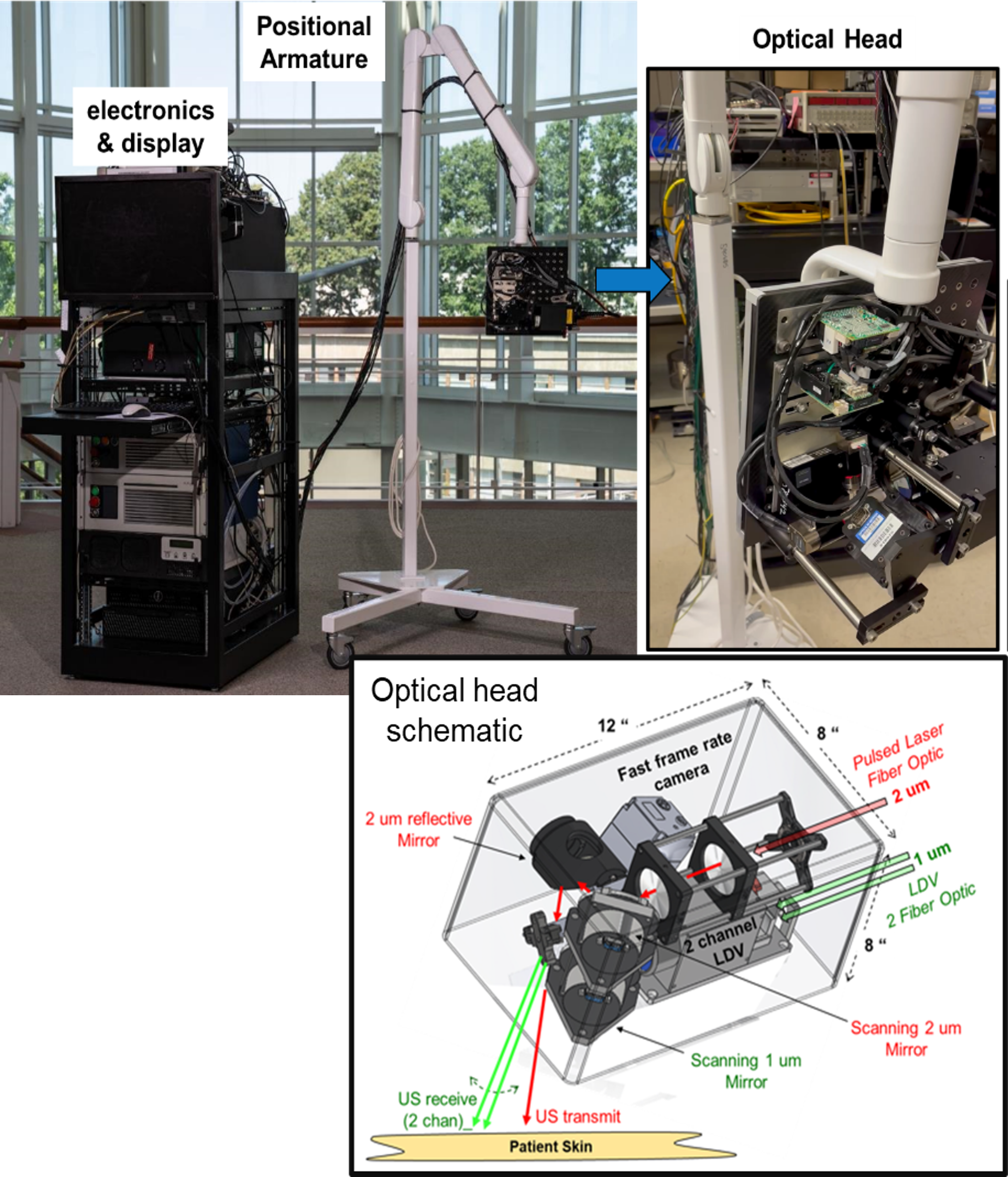
In the portable noncontact laser ultrasound system, a flexible "arm" (top left) is used to position an optical head (top right) housing a miniaturized laser source and receiver, optical fibers, mirrors, and a camera (bottom).
Motivation
Ultrasound is the most commonly used method for imaging soft tissue with many advantages: it has no harmful effects; is extremely portable, ranging from handheld devices to cart systems; and measures blood flow and elastography of tissue and organs. However, despite their size and cost, magnetic resonance imaging (MRI) and computerized tomography (CT) are the dominant technologies for tracking disease progression because they provide referenced imagery of tissue over time. Ultrasound results manually acquired at different times often vary, so an ultrasound device that can provide low-variability imagery suitable for assessing disease progression would be a welcome, innovative alternative to MRI and CT systems.
NCLUS Solution
The NCLUS system provides this breakthrough in medical imaging through its dual-laser methodology. The first laser emits short optical pulses absorbed in the skin surface that cause instantaneous thermoelastic expansion and generates ultrasound signals. The second laser, a laser Doppler vibrometer, measures the probing ultrasound echoes from the tissue interior that return to the skin surface. Any line-of-sight skin or open tissue can become a viable ultrasound source or receiver.
The system's programmable laser positioning provides a vast range of acquisition array compositions that control acoustic beam shaping and directivity for many diverse applications in medical imaging. Applications range from standard B-mode synthetic aperture imagery of soft tissue to ultrasound refraction surveys of bone and musculoskeletal injuries. Using skin fiducials, i.e., markers, NCLUS can yield a fixed reference or registered image and has the potential to offer capabilities comparable to MRI or CT. With vastly lower cost than those large imaging units, NCLUS could transform medical imaging both in and out of hospitals.
Benefits
- Noncontact acquisition overcomes significant image distortion and error caused by handheld probe manipulation. Manual probe manipulation done by different sonographers leads to significant uncertainties of measured tissue feature dimensions.
- Because the NCLUS laser scan is programmable and can provide a fixed reference frame, it can be repeated to monitor changes in tissue features over time with minimal distortion or variability and lack of reference common to results from handheld probes.
- Noncontact capability allows pain-free imaging of interior tissue through mildly to severely traumatized tissue and skin, standoff imaging of contagious patients, and imaging of body regions difficult to access.
- Automation of the acquisition scan eliminates the need for a highly skilled sonographer, thereby allowing a minimally trained operator to take the ultrasound image in nonhospital settings such as clinics and field sites.
Potential Use Cases
- Acquire ultrasound imagery through tramautized tissue, burned skin, and areas difficult to reach
- Provide registered imagery for monitoring tissue changes to track disease and injury states
- Image neonatal infants for common and serious conditions, such as bowel deterioration, in intensive care "cribs"
- Provide immersion-free, noncontact breast reflection and tomographic imaging
- Image dangerous neck and spine injuries without touching fragile body areas
- Assist in surgical procedures in real time and without interference from a contact imager
Additional Resources
U.S. Patent 10,456,044: "Systems and methods for generating non-contact ultrasound images using photoacoustic energy"
U.S. Patent 10,602,931: "System and method for non-contact ultrasound with enhanced safety"
U.S. Patent 10,835,202: "System and method for analyzing tissue using shear waves"
X. Zhang, et al., "Full Noncontact Laser Ultrasound: First Human Data," Nature, Light: Science & Applications, vol. 8, December 20, 2019.
2023 R&D 100 Award winner